
Trypanosoma brucei gambiense is a protozoan hemo-flagellate
https://pagead2.googlesyndication.com/pagead/js/adsbygoogle.js?client=ca-pub-5043583086109521Diagnosis / Pathology
Trypanosoma brucei gambiense is one of two species of trypanosoma that are
the causative agents of African sleeping sickness. Trypanosoma brucei
rhodensiense is the cause of East African sleeping sickness while Trypanosoma brucei
gambiense is the cause of West African sleeping sickness. Trypanosoma brucei
rhodensiense has a more acute presentation than Trypanosoma brucei
gambiense.
Both trypanosoma species will
present in two stages. Briefly, stage one of the infection involves the
development of a chancre lesion at the site of the vector’s (Tsetse fly) bite. Then
a haemolymphatic component ensues where the organisms can be found in the peripheral
blood and resulting in fevers, pruritus and lymphadenopathy. Though both species of
trypanosoma will result in lymphadenopathy, they differ in the location
somewhat. Trypanosoma brucei gambiense will typically produce
posterior triangular lymphadenopathy (Winterbottom’s Sign), Trypanosoma brucei
rhodensiense will produce a lymphadenopathy that is more submandibular,
axillary and inguinal.
The second stage of the infection is less acute and rapid in development with Trypanosoma brucei gambiense than Trypanosoma brucei rhodensiense. In this stage there is an invasion of the central nervous system that results in the manifestation of symptoms that give the disease its name, African sleeping sickness. Sleep disorders occur with a collection of other neuropsychiatric disorders. Severe cardiac involvement may occur. Without treatment death is the final outcome in both species infections but occurs much less rapidly in Trypanosoma brucei gambiense.
Location in the Host
Trypanosoma brucei gambiense is primarily located in the peripheral blood in the first stage and in the CSF the second stage.
Geographic Distribution
Trypanosoma brucei gambiense is endemic in West and Central Africa. Unlike Trypanosoma brucei rhodensiense where the primary reservoir is domestic cattle and wild antelope, the primary reservoir for Trypanosoma brucei gambiense is humans. Though primates and ungulates are found to carry the parasite as well.
Life Cycle

Morphology & Diagnosis
Trypanosoma brucei gambiense forms are seen in the peripheral blood in the early stage of the disease. However the trypomastigotes in the second stage are most likely seen in the cerebrospinal fluid.
The trypomastigote of Trypanosoma brucei rhodensiense and Trypanosoma brucei gambiense are morphologically indistinguishable. They stain delicately with a visible undulating membrane and flagella; a small unremarkable kinetoplast in the posterior; and a nucleus centrally located. Molecular methods can distinguish the two but many times the initial differentiation is clinically determined based on the clinical presentation and travel history of the patient.
Images
 Trypanosoma brucei gambiense in peripheral blood, Geimsa stained. Note the small kinetoplast (similar to Trypanosoma brucei rhodensiense) which when compared with the kinetoplast in the Trypanosoma cruzi trophozoite is much more large/prominent – photo courtesy of CDC/Blaine Mathison
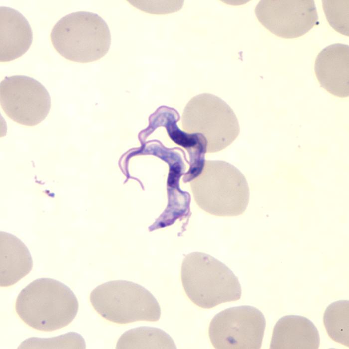